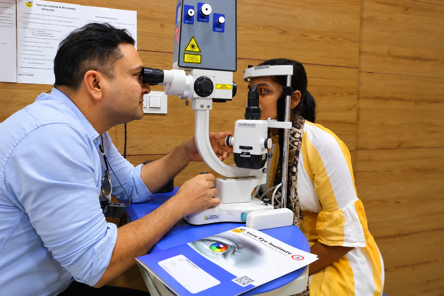
Conditions and warning signs
Retinal tears or detachment, macular conditions, retinal blood-vessel problems and bleeding inside the eye can affect vision. New flashes, a sudden shower of floaters, a curtain or shadow, or sudden distortion or loss of vision need urgent eye assessment.
Retinal detachment and tears
A retinal tear or detachment can cause flashes, new floaters or a curtain-like shadow. Depending on the findings, a specialist may discuss laser or freezing treatment for a tear, or surgery such as a scleral buckle or vitrectomy for detachment. Some operations use a gas bubble or silicone oil and require specific positioning and follow-up; the exact approach must be explained by the treating surgeon.
Macular and retinal blood-vessel conditions
Macular hole, epiretinal membrane, age-related macular degeneration, central serous chorioretinopathy and retinal vein occlusion can affect central vision in different ways. Assessment may include dilation, retinal photographs, optical coherence tomography or other tests. Depending on the diagnosis, care may involve observation, injections or a procedure; no option is suitable for everyone.
Vitreous haemorrhage and lens-related problems
Bleeding into the vitreous, a dropped lens fragment or a dislocated intraocular lens can need specialist assessment. Timing and treatment depend on the cause and how the retina is affected. Sudden vision loss needs urgent care.
Assessment and tests
An ophthalmologist may examine the retina after dilating the pupils. Depending on the symptoms, tests can include retinal photographs, optical coherence tomography (OCT) or other imaging. The doctor explains which tests are useful for your situation.
Treatment and follow-up
Some findings may be monitored; others may need treatment. Options depend on the diagnosis and may include observation, a procedure or surgery. If a procedure is proposed, ask what it involves, whether admission is needed, what recovery and positioning instructions apply, and when to return for follow-up.
Aftercare and practical questions
Follow the specific instructions from your treating team about medicines, eye drops, positioning, activity and follow-up. If a gas bubble has been placed in the eye, do not fly or travel to high altitude until the surgeon confirms it is safe. Ask for written instructions before leaving the clinic.
How the retina and vitreous work together
The retina converts incoming light into signals that travel to the brain through the optic nerve. Its central area, the macula, supports detailed activities such as reading, recognising faces and seeing fine objects. The outer retina contributes to peripheral vision. The vitreous fills much of the space inside the eye. Changes in these structures can affect central vision, side vision or the clarity of the image in different ways.
Retinal conditions are not a single diagnosis. A person with a small stable finding may need monitoring, while someone with a new tear, detachment or sudden vascular problem may need urgent assessment. Symptoms alone cannot reliably identify the cause. An examination helps establish which part of the eye is affected, whether a change is recent and how it relates to earlier records. If possible, describe when the problem began and whether it affects one eye or both.
Recognising a change that needs attention
Some longstanding floaters are harmless, but a sudden increase in floaters, new flashes, a shadow or a curtain across vision should be assessed urgently. A painless change can still be important. Do not wait for pain to develop before seeking help. Sudden loss of vision, distortion of straight lines or a new patch missing from vision also deserves timely professional assessment. A telephone description cannot replace examination of the retina.
If symptoms begin outside routine clinic hours, use an appropriate urgent eye-care or emergency service rather than waiting for a website-form reply. Avoid driving when your vision is affected. Keep the time of onset and any associated events in mind, including injury, recent surgery or a new medical problem. These details can help the clinician decide what needs to be examined first. Do not try to diagnose the condition from an online picture or another patient's experience.
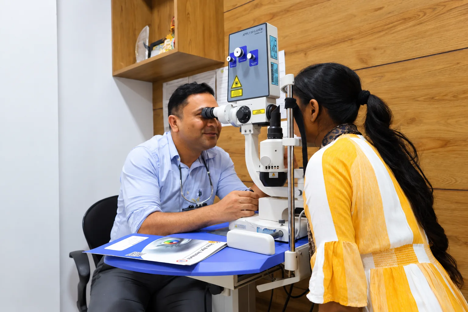
What happens during a retinal assessment
The doctor may ask about diabetes, blood pressure, previous eye operations, family history and medicines. Vision is generally assessed in each eye, followed by examination of the eye structures. Dilating drops are often used so the clinician can see the retina more fully. The effect is temporary but can make near work and bright light uncomfortable. Ask about transport and allow time for the appointment, particularly if additional tests are needed.
Imaging may help answer a specific question. Optical coherence tomography provides cross-sectional information about retinal layers; photographs document appearance; other tests may assess circulation or examine structures when the view is limited. Not everyone needs every test. Ask what each proposed investigation is intended to show, how the result will influence the plan and whether it is available at the clinic. Bring previous scans whenever possible so changes can be compared accurately.
Monitoring, procedures and individual decisions
Treatment depends on the exact condition, its location and its effect on vision. Observation can be an active management choice when the clinician considers it appropriate; it still requires a clear follow-up interval and instructions about changes to report. Other conditions may need medication, an injection, laser or surgery. The same treatment name can have different purposes in different retinal diseases, so ask how the proposed approach applies to your diagnosis.
A discussion should cover expected benefits, significant risks, alternatives and the consequences of delay or no treatment. It should also explain whether repeated visits or repeated procedures may be needed. The aim may be to preserve vision, reduce the risk of further loss or improve a particular problem; complete restoration cannot be assumed. The information on this page describes general possibilities and does not confirm that every test or procedure is offered at this clinic.
Practical planning if a procedure is recommended
Ask whether treatment is planned as an outpatient procedure, day-care surgery or an admission. The answer depends on the operation, anaesthesia and your medical needs. Let the team know about allergies, blood-thinning medicines and difficulties with transport or support at home. Do not independently stop a prescribed medicine. Your eye specialist and the doctor managing the medicine should provide instructions where a change is necessary.
Some retinal operations require specific positioning after treatment. If this is relevant, ask for a demonstration and written guidance before the operation so you can plan comfortably and safely. A gas bubble inside the eye involves important restrictions: flying and travel to higher altitude may be unsafe until the surgeon confirms otherwise. Tell any anaesthesia team about an intraocular gas bubble before another procedure. Your treating surgeon should explain the exact precautions and duration for your situation.
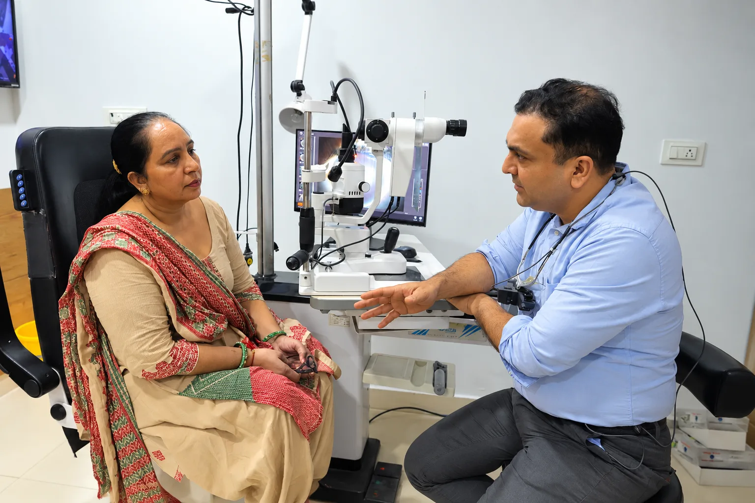
Following progress over time
Retinal care can involve more than a single appointment. The doctor may compare symptoms, visual acuity, photographs and scans over time. A stable scan does not remove the need to report a new symptom, and a small difference between scans should be interpreted by the clinician rather than in isolation. Keep follow-up dates and bring your records when changing clinics or seeking a second opinion.
Ask what changes should trigger an earlier visit and how to contact the treating team. After a procedure, worsening pain, redness or vision should be reported promptly according to the instructions given. If you cannot reach the clinic and the change is urgent, seek another appropriate emergency service. Do not replace prescribed treatment with supplements or non-prescribed eye drops on the assumption that they will repair the retina.
Supporting daily life with reduced vision
When vision remains limited, rehabilitation and practical adjustments may still help. Better lighting, appropriate magnification, larger text and changes to device settings can make specific tasks easier. Ask whether an updated spectacle prescription or low-vision assessment would be useful. Consider safety on stairs, while cooking and when crossing roads. Decisions about driving should follow professional advice and applicable legal standards, not simply how familiar a route feels.
It can be helpful to bring a relative to a complex discussion, with your permission, and to write questions beforehand. Ask for a clear diagnosis, the purpose of the next visit and a copy of important instructions. Costs, insurance paperwork and travel arrangements should be discussed before planned treatment where possible. Good communication helps you understand what is known, what remains uncertain and what you need to do next.
Describing symptoms clearly at review
Explain whether the change affects reading, faces, side vision or a particular area of the visual scene. Note whether it is present all the time and whether it began suddenly. These observations can help the clinician, but they cannot establish a diagnosis by themselves. If you have been given a specific home-monitoring method, ask how to use it and when to report a change. A home check must not delay urgent assessment or replace scheduled examinations, even if the result seems reassuring.
Frequently asked questions
Are new floaters always an emergency?
Floaters have different causes, but a sudden increase, flashes or a shadow across vision should be checked urgently.
What should I bring to an appointment?
Bring previous eye records, current prescriptions and a list of medicines and relevant medical conditions.
Will treatment restore all lost vision?
Results depend on the cause, severity and timing of care. No outcome can be promised without an individual assessment.
Will I need surgery?
That depends on the diagnosis and examination. Some retinal problems are monitored or treated without surgery.
What should I do about sudden flashes or a curtain-like shadow?
Seek urgent eye assessment. A new curtain, many floaters or sudden loss of vision should not wait for a routine appointment.
Can I travel after a retinal procedure?
Ask the surgeon before travel. If gas is placed in the eye, flying and higher-altitude travel may be unsafe until the gas has fully cleared.
Sudden vision loss or retinal warning signs should not wait for a routine appointment. This page is general information only; confirm available services with the clinic.
Request an appointmentCall the clinicWhatsApp
Clinical review and review date: confirm with the clinic before publishing this information.